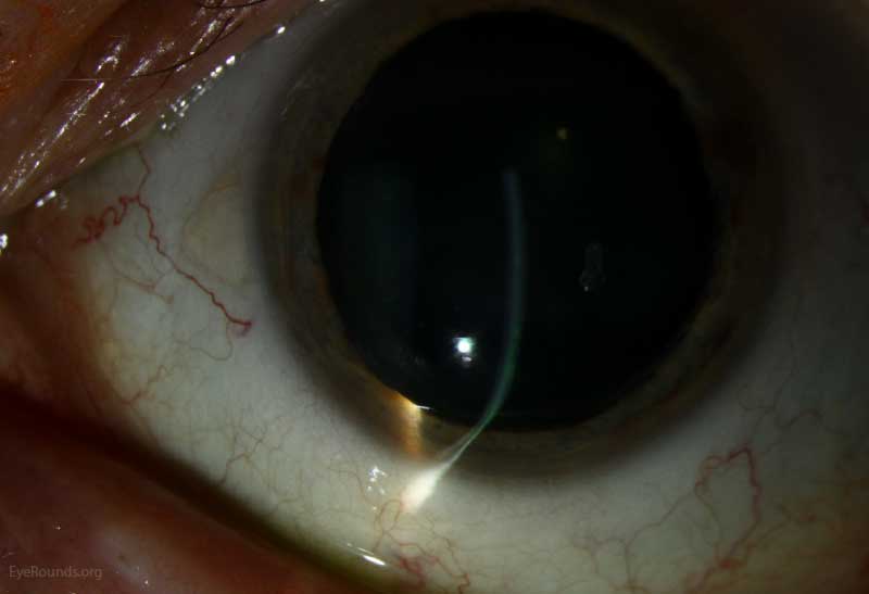
in 2004, including 116 eyes from 58 patients, 77.6% of patients were male. According to one of the large-scale studies on PMD, published by Sridhar et al. There is no known etiology for PMD yet, and no unanimous consensus whether PMD, keratoconus, and keratoglobus are distinct diseases or just different phenotypes of the same disorder. ,, Schlaeppi noticed the absence of lipid deposition, scarring, or vascularization in PMD, the so-called pellucid. , PMD manifests as impaired vision due to high against the rule and irregular astigmatism with a characteristic butterfly appearance or crab-claw pattern in corneal topography. The disease mainly affects the inferior and peripheral portions of the cornea. Pellucid marginal degeneration (PMD), initially described by Krachmer in 1978, is a bilateral noninflammatory progressive corneal ectatic disorder. Corneal cross-linking in pellucid marginal degeneration: Evaluation after five years.
#PELLUCID TOPOGRAPHY HOW TO#
How to cite this URL: Irajpour M, Noorshargh P, Peyman A. How to cite this article: Irajpour M, Noorshargh P, Peyman A. Keywords: Corneal cross-linking, Corneal ectasia, Pellucid marginal degeneration

Furthermore, regarding steep keratometry values, 25 subjects were stable, 7 had improvements, and 8 worsened.Ĭonclusion: CXL appears to be a safe and effective procedure to halt the progression of PMD. The spherical equivalent refractive error improved in 4 subjects, was stable in 25, and worsened in 11 subjects, while a 0.5 diopter or more myopic change was considered significant. Regarding the BCVA Snellen lines, 23 eyes had no significant change in pre- and postoperative examinations, 11 eyes had improvement, and 6 subjects showed worsening defined as significant when two or more lines change.

We did not find any significant difference between pre- and postoperative spherical equivalent and spherical refractive errors ( P = 0.419 and P = 0.396, respectively). No statistically significant difference was noted comparing preoperative mean logMAR best-corrected visual acuity (BCVA) and postoperative mean logMAR BCVA (0.24 ± 0.19 and 0.22 ± 0.20, P > 0.05). Results: The comparison between mean preoperative logMAR uncorrected visual acuity and 5-year postoperative evaluation revealed no significant change (1.20 ± 0.65 and 1.17 ± 0.64, P > 0.05). Visual acuity, refraction, and topography data were compared to their respective values before CXL. All subjects had undergone CXL for PMD at least 5 years before the assessments. Methods: In a retrospective study, forty eyes of forty patients were enrolled. Purpose: To evaluate the long-term outcome of corneal cross-linking (CXL) for pellucid marginal degeneration (PMD).


 0 kommentar(er)
